In a recent study performed by Vrije Universiteit Amsterdam and the University of Copenhagen, researchers used LUMICKS’ u-Flux™ laminar flow microfluidics system together with optical tweezers and fluorescence microscopy – features combined in LUMICKS’ C-Trap® – to shed light on the structural organization of mitotic chromosomes.
When preparing to undergo mitosis – a process in which replicated chromosomes are separated into two new nuclei during cell division – the genomic DNA of human cells is compacted into chromosomes approximately 5 µm long to make transport within a dividing cell easier. This chromosome compaction is driven mainly by the combined action of condensins and the enzyme topoisomerase IIα (TOP2A). Even though this dramatic chromosomal transformation has been observed using microscopy for more than a century, there is still very little known about the structural organization of mitotic chromosomes.
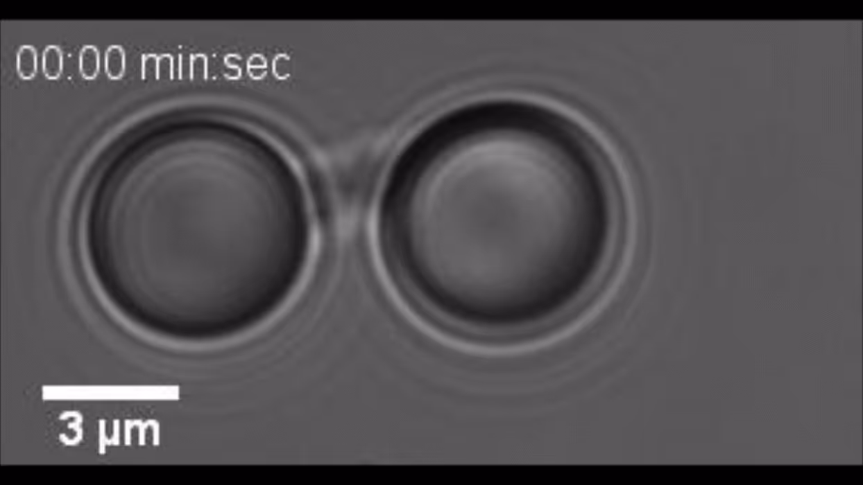
Here, the researchers used a novel workflow combining optical trapping and manipulation to extensively study mitotic chromosome mechanics and structure. This setup allows high-resolution force measurements and fluorescence visualization of chromosomes to be conducted under tightly controlled experimental conditions. By trapping and manipulating chromosomes between two beads to perform force-extension experiments, the authors found that there is a distinct non-linear stiffening of chromosomes in response to tension. Furthermore, through inducible degradation of the enzyme TOP2A specifically in mitosis, they showed that TOP2A has a role in the preservation of chromosome compaction.
This study is a great example of how LUMICKS’ dynamic single-molecule solutions can be used to directly manipulate and visualize single chromosomes to uncover the well-kept secrets of chromosome organization.
Importantly, the novel setup described here opens new avenues to a wide range of investigations that can provide great insight into the structure and dynamics of both normal and disease-associated chromosomes.
Congratulations to all the researchers from Vrije Universiteit Amsterdam and the University of Copenhagen for these exciting findings and the publication!
If you would like to find out more about their exciting results, read the full article published in Nature titled “Nonlinear mechanics of human mitotic chromosomes.“
Are you also interested in using our dynamic single-molecule solutions for your own research on chromosome organization? Do not hesitate to contact us for more information, a demo, or a quote.
Top movie courtesy of Meijering et al., Nature, 2022.
Creative Commons licence: https://creativecommons.org/licenses/by/4.0/